Foundation for maintaining high quality
The engagement in research and innovation at Quirónsalud hospitals is a basic requirement for providing the highest quality care, increasing access to the most pioneering treatments, and promoting professional development through training on state-of-the-art technology and treatments. Further, it helps to standardize and optimize processes and procedures.
Facts and figures
In 2023, 36 of our 57 hospitals were involved in scientific projects. We conducted more than 1,500 studies, 79% of which were industry-sponsored; around 6% of them were publicly funded. 11% were studies without additional funding.
The most important area of research has been oncology, the subject of approximately 55% of all clinical trials performed.
In 2023, we received a total of around €6 million in public funding (2022: around €9 million) for our clinical research activities in Spain.
Recent achievements
To optimize our clinical trial management tool, we have developed new functionalities. The tool allows the monitoring of KPIs and the development of monthly reports. Through improvements in 2023, we increased control over information registered in the platform and thus enhanced data reliability. This supports us to detect incidents and seize opportunities. Further, it is a sound basis for future decision-making.
As in previous years, we continued to make progress in actions aimed at widening research activities. We developed a project to improve clinical and organizational research management, to align research with the company’s strategy based on health, patient experience, and efficiency.
Activities carried out in 2023:
- Mapping of different subprocesses involved in clinical research
- Definition of parties involved
- Creation of multidisciplinary working groups
- Implementation of optimization measures during contract negotiation sub-processes
The project helps us to optimize and automatize processes, improve research management, quality, and results, attracts industry (which translates into more research projects), and supports talent retention.
Improving research outcomes through partnerships
Quirónsalud is part of several European research projects. This form of international collaboration allows expertise to be gathered and larger databases to be built. Projects in which we participate include:
ProCAncer-I
The project aims to address crucial questions related to prostate cancer management through the disease continuum on the one hand, and, on the other, aims to deliver a novel infrastructure enabling experimentation with AI-based solutions to improve diagnosis, treatment, and follow-up and contribute to more precise and personalized management of cancer.
For more information, click here.
PROFID
The ultimate goal is to successfully prevent the majority of the catastrophic sudden cardiac death events that occur after myocardial infarction. Thus, PROFID aims to close the gap of current clinical practice with regard to protection from sudden cardiac death after myocardial infarction.
For more information, click here.
EBRAINS
EBRAINS provides a digital research infrastructure that accelerates collaborative brain research between leading organizations and researchers across the fields of neuroscience, brain health, and brain-related technologies.
For more information, click here.
SUNRISE
As Europe continues to recover from the COVID-19 pandemic, its citizens and governments are looking ahead to future-proof society’s lifeline structures. The SUNRISE project aims to ensure greater availability, reliability, and continuity of critical infrastructures in Europe including transport, energy, water, and healthcare.
For more information, click here.
We have initiated new activities in the field of learning aimed at improving the training of our professionals in research:
- First edition of the Master’s in Clinical Research Management launched in collaboration with Universidad Europea de Madrid.
- Training program on clinical trials organized in collaboration with Roche. The program consisted of five online training modules, two of which were also delivered in person at the Hospital Universitario Quirónsalud Madrid and the Hospital Quirónsalud Barcelona.
Helium-free magnetic resonance imaging
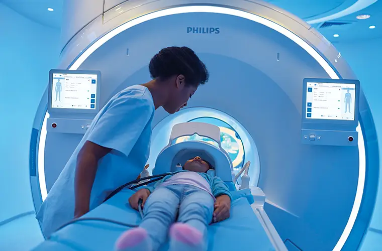
The rising cost of helium in recent years poses a risk to the sustainability of diagnostic imaging services. This chemical element is used in all magnetic resonance imaging (MRI) equipment to cool the powerful superconductors used by these machines to generate high-quality diagnostic images. However, the increasingly limited supply of a material scarce in the earth's crust and its high cost could jeopardize the normal operation of many radiology services.
A standard MRI machine needs about 1,500 liters of helium to function optimally. Generally, throughout its operating lifespan, the amount of helium in the equipment also needs to be topped up due to small gradual leaks, which poses a problem for hospital maintenance services. Moreover, the substantial economic impact becomes a secondary issue when helium refilling of an MRI is necessary and there is no availability. Should this situation occur, the equipment can be inoperative for several days, which is a cause for concern for any hospital considering the demand for these nuclear magnetic resonance examinations.
Another advantage of helium-free MRI equipment is its drastically lower weight. Conventional MRI equipment has an additional weight of close to 1.5 tons, which poses an additional engineering and construction challenge for hospitals, and even more so for hospital refurbishment when the installed base of MRI needs to be increased.
In addition, classic MRI equipment needs to have an emergency extraction system - quench tube - in case of helium gas leakage. This requirement further limits the spaces in which MRI can be installed, which is not the case with helium-free MRIs, thus facilitating accessibility to this technology in medical centers located outside hospitals.
Furthermore, the installation of helium-free MRIs also has a positive impact on the environment. Helium is a non-renewable gas and its extraction involves the drilling of oil and gas wells, which can cause environmental damage, water pollution and greenhouse gas emissions.
Aware of all these advantages, Quirónsalud was a pioneer in Spain in installing a helium-free MRI in one of its reference hospitals. Since then, 12 units have been installed in its centers.

Seeing better and safer for more reliable diagnosis
Photon Counting is a new Computed Tomography (PCTC) technology that is capable of converting each x-ray photon into an electrical signal that will be used to create the image.
The new equipment recently installed at Quirónsalud Madrid and Quirónsalud Barcelona Hospitals, are the first available in our country.
This new technology will mean an important change in the diagnosis, treatment planning and control of some pathologies.
The advantages of PCTC are many:
- Ultraspatial resolution (0.2 mm versus 0.625 mm of conventional CT). This is especially useful in the study of coronary arteries.
- Significant reduction of X-radiation dose. More than 50% in many cases. It is a key point in pediatric pathology, lung cancer screening, repetition of controls in oncological pathology.
- Reduction of metallic artifacts. This equipment minimizes the image artifacts that exist when metallic objects are present and that usually prevent a correct interpretation. In this way we can study patients with prostheses, fixation screws, implants, etc.
- Reduction of iodine contrast doses. The possibility of performing low kilovoltage monoenergetic images that highlight the visualization of iodine, as well as the minimum duration of the studies due to the extremely fast scanning speed of the equipment, make it possible to significantly reduce the amount of iodinated contrast necessary in many studies. This is important in elderly patients or those with a certain degree of renal insufficiency who are more vulnerable to the toxic effect of this type of contrast.
- Spectral imaging in all studies. The special condition of being able to measure the energy of each x-ray photon emitted allows us to characterize different tissues, i.e. we can know their composition. We can obtain different maps of materials (iodine, water, uric acid, etc.) that will help us in different diagnostic processes.
Thanks to all these characteristics, the PCTC will be a differential step in the diagnosis and follow-up of many pathological processes, with less X-ray emission and greater diagnostic reliability, fulfilling our dream of “seeing better, radiating less and characterizing better”.


